Tap again to see term. The biceps are the antagonist and the triceps are the agonist muscle.

Arms are now in extension.
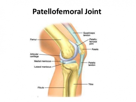
What are the articulating bones of the knee. The major ligaments in the knee joint are. Patellar ligament a continuation of the quadriceps femoris tendon distal to the patella. It attaches to the tibial.
Collateral ligaments two strap-like ligaments. They act to stabilise the hinge motion of the knee preventing. The knee joint itself manifests two formal articulations involving three bones.
The femur of the leg and the tibia of the lower leg form the primary. See full answer below. The knee joint is a synovial joint that connects three bones.
The femur tibia and patella. It is a complex hinge joint composed of two articulations. The tibiofemoral joint and patellofemoral joint.
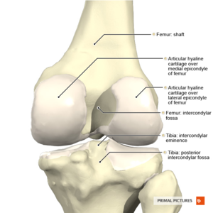
The Knee is a hinge joint made up of two bones the thigh bone femur and the shinbone tibia. There are two round knobs at the end of the femur called femoral condyles which articulate with the flat surface of the tibia called the tibial plateau. Knee joint Hinge joint.
Articulating bones are femur and tibia the patella is not classed as part of the joint nor is the fibula. Allows bending flexion and straightening extension. Muscles which move the knee are quadriceps and hamstrings.
The knee joint is the junction of three bones. The thighbone or femur. The shin bone or tibia the larger front bone of your calf.
The kneecap or patella. At its lower end the femur divides into two rounded knobs called condyles that support the bodys weight on the bone of the lower leg the tibia. The knee is the largest hinge joint in the body.
Besides flexing and extending it also rotates slightly. This movement is made possible by muscles that move the largest bones in the leg which. When knee is twisted the ligament can tear the meniscus.
The 3 Cs of knee injuries 1Cartilage. Bursa in knee joints–around patella. Water on the knee is really frictional or pre-patellar bursitis.
Talocrural or Ankle Joint. Two hinge joints involving three bones. Articular surfaces are the portions of bone that come into contact with one another within a joint.
These bony surfaces may be flush with one another as in the suture joints between the bones of the cranium or they may be separated by a cartilaginous disk as in the knee joint. The knee is a modified hinge joint a type of synovial joint which is composed of three functional compartments. The patellofemoral articulation consisting of the patella or kneecap and the patellar groove on the front of the femur through which it slides.
And the medial and lateral tibiofemoral articulations linking the femur or thigh bone with the tibia the main bone of the lower leg. The articulating bones of the knee joint are the femur tibia and fibula. Arms are now in extension.
The biceps are the antagonist and the triceps are the agonist muscle. The muscles involved in this movement are the brachialis bicep brachii brachioradialis triceps brachii and anconeus. The head of the femur articulates with the acetabulum in the pelvic bone forming the hip joint while the distal part of the femur articulates with the tibia and kneecap forming the knee joint.
By most measures the two left and right femurs are the strongest bones of the body and in humans the longest 1. Bone and Ligaments. Bones of the knee Fig1.
The longest and the heaviest bone in the bodyImportant anatomical features. Femoral head articulates with the pelvis at the acetabulum. Greater and lesser trochanters muscle attachment sites.
Linea aspera muscle attachment site. Medial and lateral epicondyles participate in the knee joint. Bones are connected to each other by joints.
The most common joint type is the. Of articulating joints which has a fibrous connective tissue capsule ligament continuous with the periosteum of the two bones and which permits a degree of freedom of movement between the two bones. The inner part of the capsule consists of the.
Knee arthroscopy is the most common type of surgical procedure performed to remove bone spurs and repair damaged cartilage in the knee joint. During the procedure a surgeon uses an arthroscope a tool about the width of a pencil with a camera and light attached to view the inside of your knee. Click card to see definition.
Tap card to see definition. Shallow joint cavity of the shoulder needs the rotator cuff muscles to help its stability Click again to see term. Tap again to see term.